
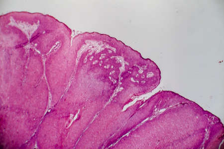
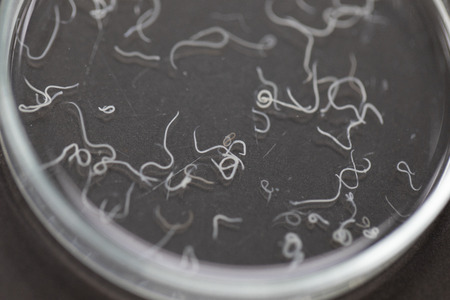
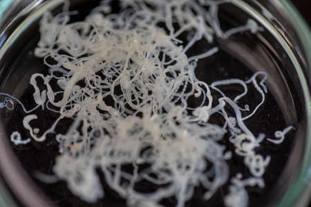
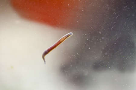
Leech on the glass. Bloodsucking animal. subclass of ringworms from the belt-type class. Hirudotherapy.
Коллекция по умолчанию
Коллекция по умолчанию
Создать новую
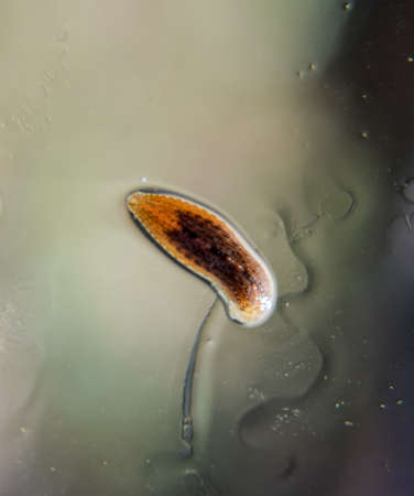
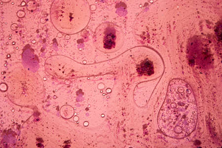
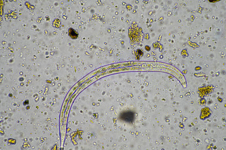
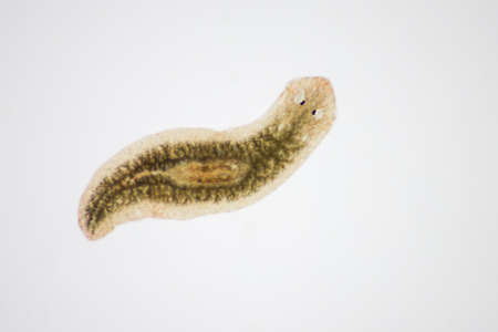
Planarian parasite (flatworm) under microscope view.
Коллекция по умолчанию
Коллекция по умолчанию
Создать новую
Extreme Close up of microscopic kidney Bowman's Capsule and Glomerulus
Коллекция по умолчанию
Коллекция по умолчанию
Создать новую
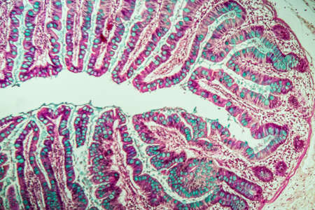
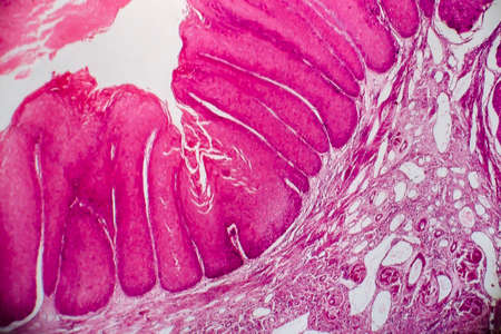
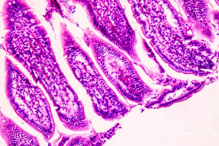
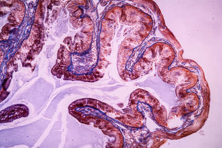
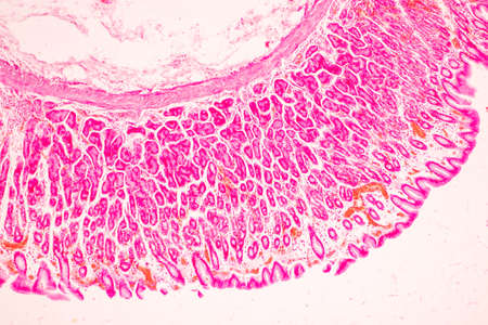
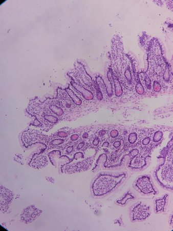
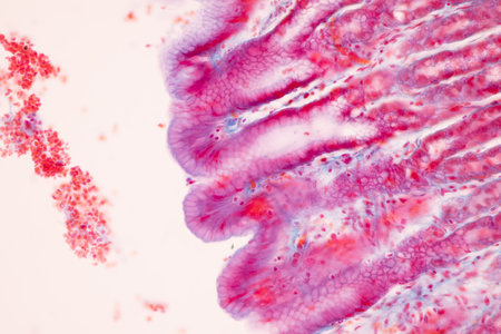
Small intestine with villi under the microscope 100x
Коллекция по умолчанию
Коллекция по умолчанию
Создать новую

Ovarian cancer, light micrograph, photo under microscope. Photograph shows a fragment of a cancerous tumor in the female ovary. Selective focus
Коллекция по умолчанию
Коллекция по умолчанию
Создать новую
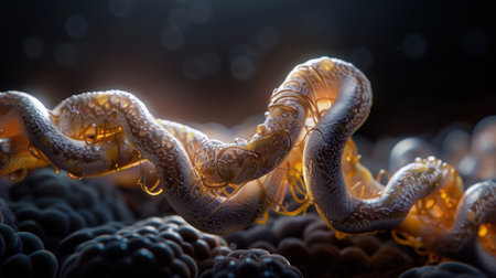
Detailed and intricate representation of snake like creature, featuring glossy texture and glowing elements, set against soft focus background of organic shapes. image evokes sense of wonder
Коллекция по умолчанию
Коллекция по умолчанию
Создать новую

Cross section of human skin under microscope view for education in laboratory.
Коллекция по умолчанию
Коллекция по умолчанию
Создать новую
Condyloma acuminatum, also known as genital warts. Light micrograph, photo under microscope
Коллекция по умолчанию
Коллекция по умолчанию
Создать новую

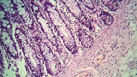
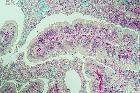
Stomach tissue under the microscope 100x
Коллекция по умолчанию
Коллекция по умолчанию
Создать новую
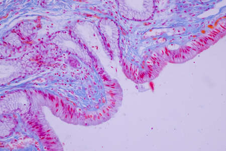
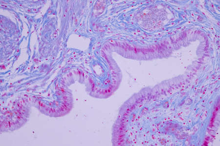
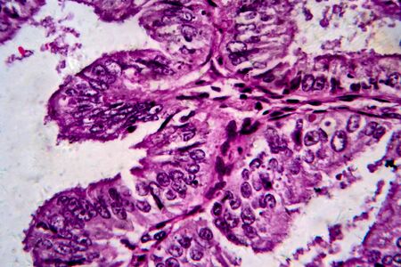
Columnar epithelium of human gall bladder under the microscope in Lab.
Коллекция по умолчанию
Коллекция по умолчанию
Создать новую
Watercolor abstract. Red paint texture of watercolor pattern or splash ink stain for design isolated on water color background
Коллекция по умолчанию
Коллекция по умолчанию
Создать новую
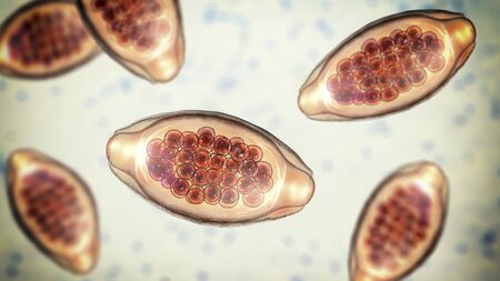
Eggs of parasitic roundworm Trichuris trichiura, or whipworm, the causative agent of trichuriasis, disease of a human large intestine, 3D illustration
Коллекция по умолчанию
Коллекция по умолчанию
Создать новую
Soothing Pink Abstract background with little impurities
Коллекция по умолчанию
Коллекция по умолчанию
Создать новую
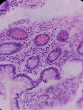
Education anatomy and Histological sample of Human under the microscope.
Коллекция по умолчанию
Коллекция по умолчанию
Создать новую
Rare image of Ghost flatworm - Maricola (Planarian) triclad flatworms in reef aquarium glass
Коллекция по умолчанию
Коллекция по умолчанию
Создать новую

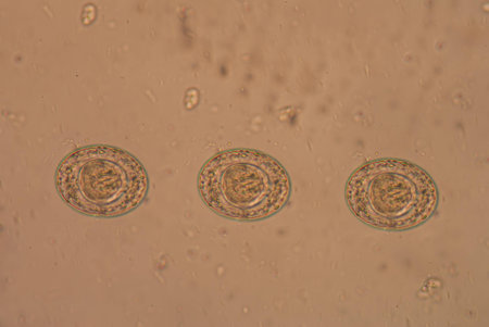
Egg of Ascaris lumbricoides (roundworm) in human stool, analyze by microscope, 400x
Коллекция по умолчанию
Коллекция по умолчанию
Создать новую

Egg of parasite in stool examition testing finding with microscope.
Коллекция по умолчанию
Коллекция по умолчанию
Создать новую
Condyloma acuminatum, also known as genital warts. Light micrograph, photo under microscope
Коллекция по умолчанию
Коллекция по умолчанию
Создать новую

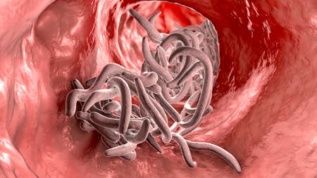
Parasitic worms in human small intestine, 3D illustration. Ascaris lumbricoides and other round worms
Коллекция по умолчанию
Коллекция по умолчанию
Создать новую
Stomach tissue under the microscope 100x
Коллекция по умолчанию
Коллекция по умолчанию
Создать новую
Spaghetti in motion, flying noodles in air. suitable for food, noodle or spaghetti themes
Коллекция по умолчанию
Коллекция по умолчанию
Создать новую
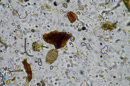
Dive into a stunning microscopic view showcasing vibrant microorganisms in an aquatic setting. This image reveals the intricacies of diverse life forms through scientific observation.
Коллекция по умолчанию
Коллекция по умолчанию
Создать новую
Tissue of Stomach Human under the microscope in Lab.
Коллекция по умолчанию
Коллекция по умолчанию
Создать новую
A striking image of a colorful purple worm set against a vibrant yellow background. Ideal for creative projects, educational materials, or graphic design.
Коллекция по умолчанию
Коллекция по умолчанию
Создать новую
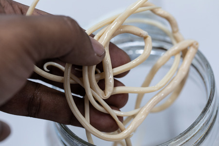
Ascariasis is a disease caused by the parasitic roundworm Ascaris lumbricoides for education in laboratories.
Коллекция по умолчанию
Коллекция по умолчанию
Создать новую
Human egg cell, 3D illustration closeup
Коллекция по умолчанию
Коллекция по умолчанию
Создать новую

Columnar epithelium of human gall bladder under the microscope in Lab.
Коллекция по умолчанию
Коллекция по умолчанию
Создать новую

Parasitic worms in the lumen of intestine, 3D illustration. Ascaris lumbricoides, Enterobius vermicularis, and other round worms
Коллекция по умолчанию
Коллекция по умолчанию
Создать новую
Ascariasis is a disease caused by the parasitic roundworm Ascaris lumbricoides for education in laboratories.
Коллекция по умолчанию
Коллекция по умолчанию
Создать новую
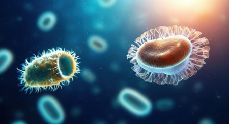
Bacteria cells under microscope, 3D illustration.
Коллекция по умолчанию
Коллекция по умолчанию
Создать новую

Histological Uterus human, Uterine tube human, Placenta human and Umbilical cord Human under the microscope for education.
Коллекция по умолчанию
Коллекция по умолчанию
Создать новую

Fusobacterium, 3D illustration. An oral bacterium, causes periodontal diseases, periodontal plague formation, sore throat, Lemmieres syndrome. It is also associated with preterm birth and colon cancer
Коллекция по умолчанию
Коллекция по умолчанию
Создать новую
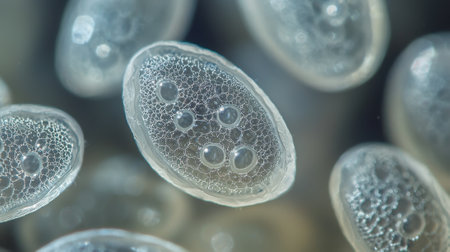
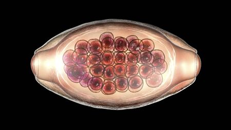
Ascaris lumbricoides, a large roundworm, unfertilized egg, 3D illustration
Коллекция по умолчанию
Коллекция по умолчанию
Создать новую
Ascariasis is a disease caused by the parasitic roundworm Ascaris lumbricoides for education in laboratories.
Коллекция по умолчанию
Коллекция по умолчанию
Создать новую

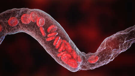
Blood vessel with flowing blood cells, 3D illustration. Small blood vessels, capillaries
Коллекция по умолчанию
Коллекция по умолчанию
Создать новую

microorganisms and soil biology, with nematodes and fungi under the microscope in a soil compost sample
Коллекция по умолчанию
Коллекция по умолчанию
Создать новую
Histopathology of cholera under microscope view for education.
Коллекция по умолчанию
Коллекция по умолчанию
Создать новую
Schistosoma is a genus of trematodes
Коллекция по умолчанию
Коллекция по умолчанию
Создать новую

Columnar epithelium of human gall bladder under the microscope in Lab.
Коллекция по умолчанию
Коллекция по умолчанию
Создать новую
The study of Tapeworm infection is caused by ingesting food or water contaminated with tapeworm eggs or larvae in laboratory.
Коллекция по умолчанию
Коллекция по умолчанию
Создать новую

Fusobacterium, 3D illustration. An oral bacterium, causes periodontal diseases, periodontal plague formation, sore throat, Lemmieres syndrome. It is also associated with preterm birth and colon cancer
Коллекция по умолчанию
Коллекция по умолчанию
Создать новую
This 3D illustration shows a peptic ulcer affecting the stomach surface with visible tissue damage and surrounding elements captured in detail.
Коллекция по умолчанию
Коллекция по умолчанию
Создать новую
Trumpet animal as a microscopic plankton animal in drops of water
Коллекция по умолчанию
Коллекция по умолчанию
Создать новую
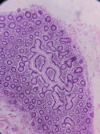
Characteristics of Lichen, hyphae and Symbiotic algae under the microscope for education.
Коллекция по умолчанию
Коллекция по умолчанию
Создать новую
Taenia is a genus of tapeworm (a type of helminth) that includes some important parasites of livestock.
Коллекция по умолчанию
Коллекция по умолчанию
Создать новую
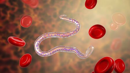
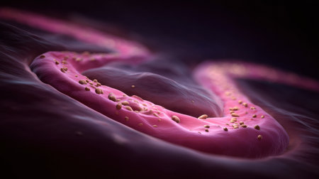
Wuchereria bancrofti, a roundworm nematode, one of the causative agents of lymphatic filariasis, 3D illustration showing presence of sheath around the worm and tail nuclei non-extending to tip
Коллекция по умолчанию
Коллекция по умолчанию
Создать новую
Signet ring cell carcinoma of the stomach, light micrograph, photo under microscope
Коллекция по умолчанию
Коллекция по умолчанию
Создать новую
Bacillary dysentery, light micrograph, photo under microscope showing presence of bacteria and accumulation of inflammatory cells in intestinal epithelium
Коллекция по умолчанию
Коллекция по умолчанию
Создать новую
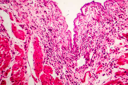
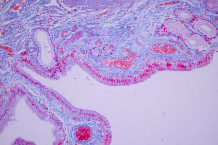
Histopathology of human under microscope view for education in laboratory.
Коллекция по умолчанию
Коллекция по умолчанию
Создать новую

A deep stunning-sea siphonophore drifting gracefully in the ocean depths
Коллекция по умолчанию
Коллекция по умолчанию
Создать новую

The human whipworm (Trichuris trichiura or Trichocephalus trichiuris) is a round worm (a type of helminth) that causes trichuriasis (a type of helminthiasis which is one of the neglected tropical diseases) when it infects a human large intestine.
Коллекция по умолчанию
Коллекция по умолчанию
Создать новую

Education anatomy and Histological sample Spinal cord Tissue under the microscope.
Коллекция по умолчанию
Коллекция по умолчанию
Создать новую
bacteria under the microscope background, background bacteria
Коллекция по умолчанию
Коллекция по умолчанию
Создать новую

Endometriosis, a disorder in which cells similar to those in the endometrium grow outside the uterus. Light micrograph, photo under microscope
Коллекция по умолчанию
Коллекция по умолчанию
Создать новую
Microscopic Examination of the Sclerotized Cephalopharyngeal Skeleton: The Powerful Internal Mouthparts of a Spiral Fly Larva
Коллекция по умолчанию
Коллекция по умолчанию
Создать новую
Close-up view of glowing bacteria and viruses with spiky exteriors, floating in a dark blue, luminous, and abstract background.
Коллекция по умолчанию
Коллекция по умолчанию
Создать новую
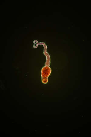
Billharzia pathogen larva Furcozerkarien 200x
Коллекция по умолчанию
Коллекция по умолчанию
Создать новую
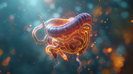
An illustration of the small colon, duodenum, stomach, intestine, and digestive tract
Коллекция по умолчанию
Коллекция по умолчанию
Создать новую
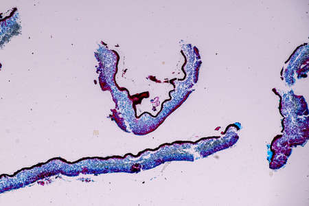
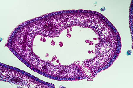
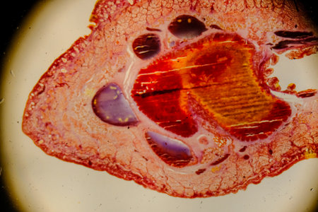
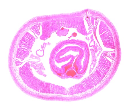
Slow worm histology bowel transverse 100x
Коллекция по умолчанию
Коллекция по умолчанию
Создать новую
Heather leaf cross section under the microscope, 200x
Коллекция по умолчанию
Коллекция по умолчанию
Создать новую
Columnar epithelium of human gall bladder under the microscope in Lab.
Коллекция по умолчанию
Коллекция по умолчанию
Создать новую
Pancreas cancer cell under microscope view for medical education.
Коллекция по умолчанию
Коллекция по умолчанию
Создать новую
Ascariasis is a disease caused by the parasitic roundworm Ascaris lumbricoides for education in laboratories.
Коллекция по умолчанию
Коллекция по умолчанию
Создать новую
Histopathology of human liver under microscope view for medical education.
Коллекция по умолчанию
Коллекция по умолчанию
Создать новую

medical microscopy animal parasiteras schistosome blood flukes
Коллекция по умолчанию
Коллекция по умолчанию
Создать новую
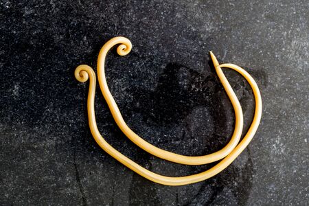
Ascariasis is a disease caused by the parasitic roundworm Ascaris lumbricoides for education in laboratories.
Коллекция по умолчанию
Коллекция по умолчанию
Создать новую
A close up of many small, round, clear objects with a lot of holes in them. The objects are all different sizes and are scattered throughout the image. Scene is one of curiosity and wonder
Коллекция по умолчанию
Коллекция по умолчанию
Создать новую
Parasitic worms in the lumen of intestine, 3D illustration. Ascaris lumbricoides, Enterobius vermicularis, and other round worms
Коллекция по умолчанию
Коллекция по умолчанию
Создать новую

Histopathology of human under microscope view for education in laboratory.
Коллекция по умолчанию
Коллекция по умолчанию
Создать новую
fungal hyphae and soil fungi in a soil sample, showing the living soil form a farm
Коллекция по умолчанию
Коллекция по умолчанию
Создать новую

Abstract science background- pyloric division of the stomach of the dog. Cell biology
Коллекция по умолчанию
Коллекция по умолчанию
Создать новую
Columnar epithelium of human gall bladder under the microscope in Lab.
Коллекция по умолчанию
Коллекция по умолчанию
Создать новую
Pancreas cancer cells, light micrograph for medical education.
Коллекция по умолчанию
Коллекция по умолчанию
Создать новую
Vasoconstriction, clot formation in blood vessel. Erythrocytes flowing through narrowing blood vessel, 3D illustration
Коллекция по умолчанию
Коллекция по умолчанию
Создать новую
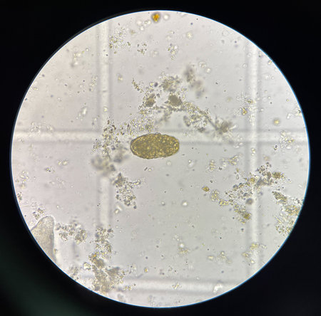
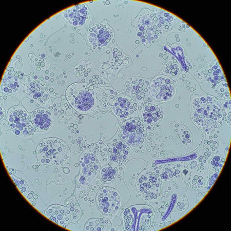
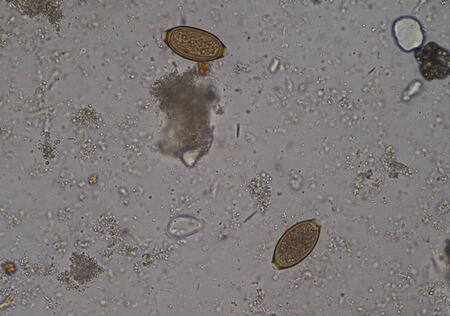
human parasite in stool examination test.
Коллекция по умолчанию
Коллекция по умолчанию
Создать новую
A microscopic image shows numerous budding yeast cells, likely Saccharomyces cerevisiae, under magnification. The circular and oval cells are seen in various stages of division, representing microbiology, fermentation, scientific research, and biology.
Коллекция по умолчанию
Коллекция по умолчанию
Создать новую
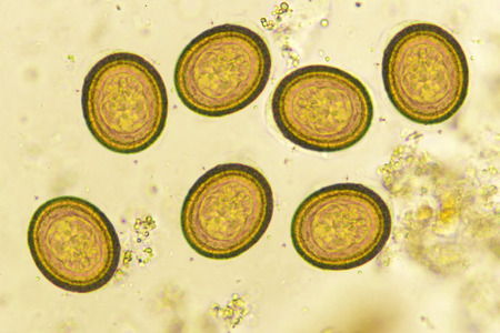
Eggs of Taenia in stool, analyze by microscope
Коллекция по умолчанию
Коллекция по умолчанию
Создать новую
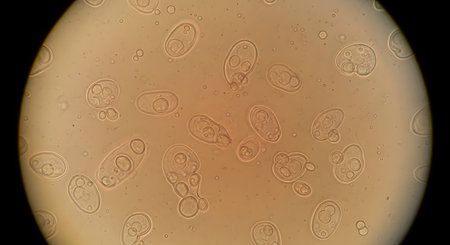
Egg of parasite in stool examition testing finding with microscope.
Коллекция по умолчанию
Коллекция по умолчанию
Создать новую
Leech cross section showing internal anatomical structures stained
Коллекция по умолчанию
Коллекция по умолчанию
Создать новую
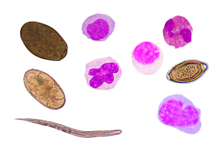
blood cells and parasite eggs on white background.
Коллекция по умолчанию
Коллекция по умолчанию
Создать новую
Egg of parasitic roundworm Trichuris trichiura, or whipworm, the causative agent of trichuriasis, disease of a human large intestine, 3D illustration
Коллекция по умолчанию
Коллекция по умолчанию
Создать новую
microorganisms and soil biology, with nematodes and fungi under the microscope. in a soil and compost sample
Коллекция по умолчанию
Коллекция по умолчанию
Создать новую
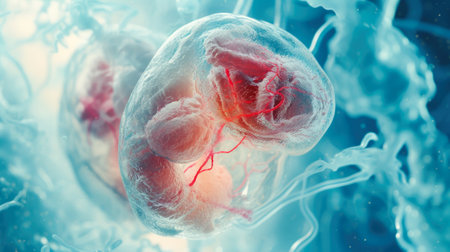
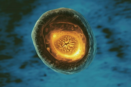
Embryonic Development: Microscopic View of a Translucent Embryo
Коллекция по умолчанию
Коллекция по умолчанию
Создать новую

Abstract macro image of particles looking like bacteria, macro shot, microbiology theme
Коллекция по умолчанию
Коллекция по умолчанию
Создать новую

Three-dimensional wishing model of human intestines on a blue background, 3D rendering
Коллекция по умолчанию
Коллекция по умолчанию
Создать новую
Earthworm under the microscope, background Lumbricidae
Коллекция по умолчанию
Коллекция по умолчанию
Создать новую

The malaria-infected red blood cells. 3D illustration showing malaria parasite Plasmodium falciparum in schizont stage inside red blood cells, the causative agent of tropical malaria
Коллекция по умолчанию
Коллекция по умолчанию
Создать новую
Endometrial adenocarcinoma, light micrograph, photo under microscope
Коллекция по умолчанию
Коллекция по умолчанию
Создать новую
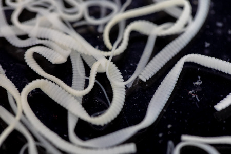
The study parasite or worms is a freshwater fish parasite in laboratory for education.
Коллекция по умолчанию
Коллекция по умолчанию
Создать новую
3D illustration of abstract light background with bokeh defocused lights
Коллекция по умолчанию
Коллекция по умолчанию
Создать новую
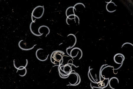
science aquaculture fish parasite hook clip worm micrograph
Коллекция по умолчанию
Коллекция по умолчанию
Создать новую
Macro view of colorful abstract texture, resembling organic biological forms. Bright pink and red hues create a dynamic and fluid pattern.
Коллекция по умолчанию
Коллекция по умолчанию
Создать новую
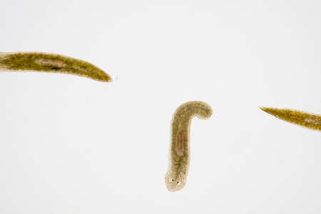
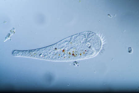
Planarian parasite (flatworm) under microscope view.
Коллекция по умолчанию
Коллекция по умолчанию
Создать новую

Colon polyp, one of the largest polyps
Коллекция по умолчанию
Коллекция по умолчанию
Создать новую
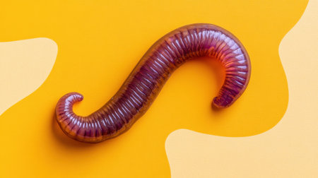
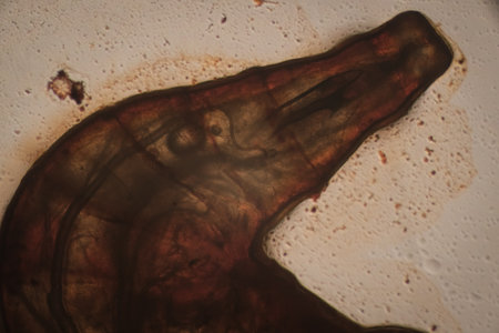
A red-brown suction worm in an aquarium on the disk.
Коллекция по умолчанию
Коллекция по умолчанию
Создать новую
3d illustration of abstract background with glowing multicolor spheres in space
Коллекция по умолчанию
Коллекция по умолчанию
Создать новую
Showing Light micrograph of the Trachea, Thymus, Parathyroid gland and Tonsil human under the microscope for education in the laboratory.
Коллекция по умолчанию
Коллекция по умолчанию
Создать новую

Histopathology of interstitial nephritis, light micrograph, photo under microscope
Коллекция по умолчанию
Коллекция по умолчанию
Создать новую

Parasitic worms in the lumen of intestine, 3D illustration. Growth and multiplication of nematode worms invading human intestine
Коллекция по умолчанию
Коллекция по умолчанию
Создать новую
Cytomegalovirus CMV in a human cell, owl's eye inclusion in nucleus, multinucleated cell, 3D illustration. It is herpes virus, causes diseases in fetus, organ transplant patients, HIV infected people
Коллекция по умолчанию
Коллекция по умолчанию
Создать новую
Legion-Media
Создайте свои проекты на основе качественных стоковых фотографий и видео.


Copyright © Legion-Media.